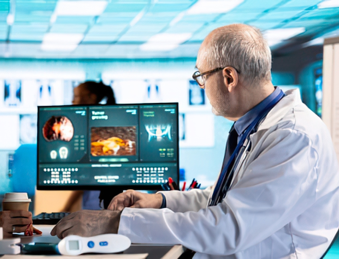
Medical imaging used to mean one thing: a radiologist, a lightbox (or today, a monitor), and a trained eye scanning scores of scans for subtle signs of disease. That process still exists, but it no longer works alone. Artificial intelligence now reads alongside radiologists in thousands of hospitals worldwide, flagging strokes before a human even opens the file, catching lung nodules a tired eye might miss at 2 a.m., and compressing MRI scan times that used to keep patients in the machine for 45 minutes.
This shift is not experimental anymore. As of early 2026, the FDA has authorized more than 1,500 AI-enabled medical devices, and radiology accounts for roughly three-quarters of them. That number has been climbing fast from an average of about 21 new clearances a month in 2024 to roughly 30 a month by mid-2026. AI in medical imaging has moved from a research curiosity to core clinical infrastructure.
This guide breaks down what AI-powered medical imaging actually is, how it works under the hood, where it’s already changing patient outcomes, and what to watch out for before adopting it.
Key Takeaways
- AI-powered medical imaging uses machine learning mainly deep learning and computer vision to help detect, measure, and prioritize abnormalities in scans like X-rays, CT, MRI, ultrasound, and mammography.
- It works primarily as a “second reader” or triage tool that flags urgent cases and reduces radiologist workload, not as a replacement for physicians.
- The FDA has cleared more than 1,500 AI-enabled medical devices, with radiology making up around 76% of the total as of 2026.
- Proven use cases include stroke detection (large vessel occlusion), lung nodule and cancer screening, pulmonary embolism triage, diabetic retinopathy screening, and mammography support.
- Real-world performance can drop meaningfully outside the hospitals where a model was trained; validation and monitoring matter as much as the initial accuracy claim.
- Successful adoption depends on data infrastructure, workflow integration (PACS/RIS), and ongoing model monitoring, not just picking a tool with a good headline accuracy score.
AI-Powered Medical Imaging: What Is It?
AI-driven medical imaging is the application of artificial intelligence primarily deep learning models built on convolutional neural networks (CNNs) and, increasingly, transformer-based and foundation models to analyze diagnostic images such as X-rays, CT scans, MRIs, ultrasounds, and mammograms.
Instead of a radiologist manually reviewing every pixel of every scan, an AI model is trained on large datasets of labeled medical images to recognize visual patterns associated with specific conditions: a hairline fracture, a blocked artery, a suspicious mass, early signs of diabetic retinopathy. The model then applies that pattern recognition to new, unseen scans, usually in seconds, and outputs a finding, a probability score, a segmentation (an outline of the area of concern), or a triage flag for urgent review.
It’s important to be precise about what these systems do and don’t do. Nearly all FDA-cleared imaging AI tools are classified as assistive or concurrent reading aids; they support a licensed clinician’s judgment rather than issuing an independent, unsupervised diagnosis. The clinician continues to be the official decision-maker.
How AI-Powered Medical Imaging Differs From Traditional Image Analysis
Traditional computer-aided detection (CAD) software, common since the early 2000s, relied on hand-engineered rules: if a shape had certain dimensions or density, flag it. These systems were rigid and produced a lot of false positives.
Modern AI-powered imaging tools learn patterns directly from data rather than following fixed rules. This makes them far more adaptable; a single AI-powered medical imaging task uses tens of findings across an entire CT scan, something rule-based CAD could never do efficiently.
How AI-Powered Medical Imaging Actually Works
At a high level, most clinical imaging AI systems follow the same pipeline:
1. Image Acquisition and Ingestion
The scan is captured by standard imaging hardware (CT, MRI, X-ray, ultrasound, PET) and stored in DICOM format, then routed usually automatically from the hospital’s PACS (Picture Archiving and Communication System) to the AI engine.
2. Preprocessing
The raw image is normalized: resolution standardized, noise reduced, and, for 3D modalities like CT and MRI, the volume is reconstructed into slices the model can process.
3. Model Inference
A trained neural network analyzes the image. Depending on the task, it may perform:
- Classification — determining whether a finding is present or absent (e.g., pneumonia: yes/no)
- Detection — locating and bounding a specific abnormality (e.g., a lung nodule)
- Segmentation — outlining the precise boundary of a structure (e.g., a tumor volume for radiation planning)
- Triage/prioritization — reordering the radiologist’s worklist so the most urgent case (e.g., a suspected stroke) appears first
4. Clinical Output and Integration
Results are pushed back into the PACS or radiology reporting software as an overlay, a flag, or a structured report, so radiologists see the AI’s findings directly inside their normal workflow rather than in a separate application.
5. Human Review and Sign-Off
A licensed radiologist or physician confirms, adjusts, or overrides the AI output before it becomes part of the patient’s official record. This human-in-the-loop step is central to how these tools are regulated and used today.
Benefits of AI in Medical Imaging
- Faster time-to-diagnosis for time-critical conditions like stroke and PE, where minutes directly affect outcomes
- Reduced radiologist workload by automating routine measurements, pre-screening normal studies, and prioritizing urgent cases
- More consistent second-read coverage, especially valuable in high-volume screening programs and under-resourced facilities
- Expanded access to specialist-level screening in primary care and rural settings, such as retinopathy screening without an on-site ophthalmologist
- Quantitative, reproducible measurements (tumor volume, vessel diameter, nodule size over time) that reduce inter-reader variability
Limitations and Risks to Understand
- Performance drop outside the training environment. Models validated at one institution can lose significant specificity in some studies up to 20-25 percentage points when deployed at a different hospital with different scanners, patient populations, or protocols. Site-specific validation matters.
- Not a replacement for clinical judgment. Virtually all cleared tools are assistive, not autonomous. Final diagnostic responsibility stays with the treating clinician.
- Data and integration overhead. AI tools need a modern PACS/RIS environment and reliable DICOM routing to work well; legacy imaging infrastructure can blunt or block the benefit entirely.
- Regulatory and liability questions. Clearance pathways (510(k) vs. De Novo), post-market monitoring, and medico-legal responsibility for AI-assisted misses are still evolving areas that healthcare organizations need to track.
- Bias and generalizability. Training data that underrepresents certain demographics, scanner types, or disease presentations can produce uneven accuracy across patient populations a key reason transparent clinical validation data matters when evaluating a vendor.
How Healthcare Organizations Can Evaluate and Adopt Imaging AI
- Start with the clinical problem, not the technology. Identify a specific bottleneck missed nodules, slow stroke triage, screening backlog before shopping for a tool.
- Demand transparent clinical validation data. Ask for multi-site study results, not just a single-institution accuracy figure, and check whether the evidence has been peer-reviewed or published.
- Assess integration effort honestly. Confirm the tool works with your existing PACS/RIS and DICOM routing before committing, since poor integration is one of the most common reasons AI tools go unused after purchase.
- Monitoring plan, not just deployment. Model performance can drift over time as patient populations, scanners, or protocols change; build in a process to re-validate periodically.
- Involve radiologists and clinicians early. Tools that get folded seamlessly into existing reading workflows see far higher adoption than those bolted on as a separate step.
- Build the underlying data pipeline first. Reliable, well-structured medical imaging data clean DICOM metadata, proper labeling, and interoperable storage is the foundation every imaging AI model depends on. Without it, even the best algorithm underperforms.
Final Thoughts
AI-powered medical imaging isn’t a future technology anymore; it’s already embedded in stroke pathways, lung cancer screening programs, and mammography suites at thousands of hospitals. The organizations getting the most value from it aren’t necessarily the ones with the flashiest algorithm; they’re the ones with clean, well-structured imaging data, solid systems integration, and a clear clinical workflow for putting AI output in front of the right clinician at the right moment.
That last part the data foundation underneath the algorithm is where most imaging AI initiatives actually succeed or fail.
Building or evaluating an AI-driven imaging or diagnostics initiative? Deep Data Insight helps healthcare and life sciences teams design the data infrastructure, model integration, and AI/ML systems that make imaging AI reliable in real clinical environments. Talk to our team about your project.
FAQs
Is AI-powered medical imaging the same as a diagnosis from a doctor?
No. Nearly all FDA-cleared imaging AI tools are assistive or concurrent reading aids. They flag findings, prioritize cases, or provide measurements, but a licensed radiologist or physician reviews and confirms the result before it becomes part of a patient’s official diagnosis.
How accurate is AI in medical imaging?
Accuracy varies by task and vendor, but leading tools in categories like stroke and pulmonary embolism detection report sensitivity and specificity in the mid-to-high 90% range in controlled studies. Real-world accuracy at a specific hospital can be lower than published figures, since performance depends heavily on how similar that hospital’s scanners and patient population are to the AI’s training data.
Will AI replace radiologists?
Most evidence points the other way: AI is expanding radiologist capacity rather than replacing the role. Demand for radiologists remains high, and current tools are designed to reduce workload and catch misses, not to operate independently of a qualified physician.
Which imaging modalities does AI work with?
AI models are in clinical use across CT, MRI, X-ray, ultrasound, mammography, and retinal imaging, with CT and X-ray currently having the largest number of FDA-cleared applications.
What is the FDA’s role in approving medical imaging AI?
The FDA clears AI-enabled medical devices primarily through the 510(k) pathway (showing substantial equivalence to an existing device) or the De Novo pathway (for novel device types). As of 2026, radiology represents the large majority of all FDA-cleared AI-enabled medical devices.
What’s needed to implement AI imaging tools in a hospital or clinic?
At minimum: a compatible PACS/RIS environment, reliable DICOM data routing, IT support for integration and monitoring, and a clinical workflow plan for how radiologists will review and act on AI output.